WELCOME TO OUR PARKINSON'S PLACE!

I HAVE PARKINSON'S DISEASES AND THOUGHT IT WOULD BE NICE TO HAVE A PLACE WHERE THE CONTENTS OF UPDATED NEWS IS FOUND IN ONE PLACE. THAT IS WHY I BEGAN THIS BLOG.

I COPY NEWS ARTICLES PERTAINING TO RESEARCH, NEWS AND INFORMATION FOR PARKINSON'S DISEASE, DEMENTIA, THE BRAIN, DEPRESSION AND PARKINSON'S WITH DYSTONIA. I ALSO POST ABOUT FUNDRAISING FOR PARKINSON'S DISEASE AND EVENTS. I TRY TO BE UP-TO-DATE AS POSSIBLE.

I AM NOT RESPONSIBLE FOR IT'S CONTENTS. I AM JUST A COPIER OF INFORMATION SEARCHED ON THE COMPUTER. PLEASE UNDERSTAND THE COPIES ARE JUST THAT, COPIES AND AT TIMES, I AM UNABLE TO ENLARGE THE WORDING OR KEEP IT UNIFORMED AS I WISH. IT IS IMPORTANT TO UNDERSTAND I AM A PERSON WITH PARKINSON'S DISEASE. I HAVE NO MEDICAL EDUCATION,

I JUST WANT TO SHARE WITH YOU WHAT I READ ON THE INTERNET. IT IS UP TO YOU TO DECIDE WHETHER TO READ IT AND TALK IT OVER WITH YOUR DOCTOR. I AM JUST THE COPIER OF DOCUMENTS FROM THE COMPUTER. I DO NOT HAVE PROOF OF FACT OR FICTION OF THE ARTICLE. I ALSO TRY TO PLACE A LINK AT THE BOTTOM OF EACH ARTICLE TO SHOW WHERE I RECEIVED THE INFORMATION SO THAT YOU MAY WANT TO VISIT THEIR SITE.

THIS IS FOR YOU TO READ AND TO ALWAYS KEEP AN OPEN MIND.

PLEASE DISCUSS THIS WITH YOUR DOCTOR, SHOULD YOU HAVE ANY QUESTIONS, OR CONCERNS. NEVER DO ANYTHING WITHOUT TALKING TO YOUR DOCTOR FIRST..

I DO NOT MAKE ANY MONEY FROM THIS WEBSITE. I VOLUNTEER MY TIME TO HELP ALL OF US TO BE INFORMED.

I WILL NOT ACCEPT ANY ADVERTISEMENT OR HEALING POWERS, HEALING FROM HERBS AND ETC. UNLESS IT HAS GONE THROUGH TRIALS AND APPROVED BY FDA. IT WILL GO INTO SPAM.

THIS IS A FREE SITE FOR ALL WITH NO ADVERTISEMENTS

THANK YOU FOR VISITING! TOGETHER WE CAN MAKE A DIFFERENCE!

TRANSLATE

Thursday, September 24, 2015

NEW TECHNIQUE NEGOTIATES NEURON JUNGLE TO TARGET SOURCE OF PARKINSON'S DISEASE


23 September 2015London, UK
 
Newcastle University
Imperial College London

Press release

Researchers from Imperial College London and Newcastle University believe they have found a potential new way to target cells of the brain affected by Parkinson's disease.
The new technique is relatively non-invasive and has worked to improve symptoms of the disease in rats.
Parkinson's disease causes progressive problems with movement,posture and balance. It is currently treated with drugs, but these have severe side-effects and can become ineffective after around five years. The only treatment subsequently available to patients is deep brain stimulation, a surgical technique where an electrical current is used to stimulate nerve cells in the brain.
As well as being an invasive treatment, it has mixed results - some patients benefit while others experience no improvement or even deteriorate. Researchers believe this is because the treatment is imprecise, stimulating all types of nerve cells, not just the intended target.
The new study, published in the journal Molecular Neurodegeneration, examined a less invasive and more precise alternative, designed to target and stimulate a particular type of nerve cell called cholinergic neurons. These are found within a part of the brain called the pedunculopontine nucleus, or PPN.

"This paper will help us understand how deep-brain stimulation works, but more importantly it is a step towards offering less invasive treatment options to patients with Parkinson's and other neurodegenerative disorders."  

Dr Joanna Elson, at the
Institute of Genetic Medicine at Newcastle University
"If you were to peer inside the PPN, it is like a jungle with a massive variety of nerve cells that behave differently and have different jobs to do," said Dr Ilse Pienaar, Honorary Lecturer in Neuroscience at Imperial College London
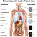
Scientists already suspect that cholinergic neuron cells are involved in Parkinson's disease. This is because in post mortem studies of patients? brains, about half of these cells have perished, for reasons that are currently unknown.
The researchers from Imperial College London and Newcastle University worked with rats that had been treated to recreate the symptoms of Parkinson's disease. They used a harmless virus to deliver a specially-designed genetic 'switch' to the cholinergic neurons. The rats were then given a drug that was designed to activate the 'switch' and stimulate the target neurons.
Following the treatment the rats made an almost complete recovery and were able to move normally.
Dr Pienaar adds: "This study confirms that cholinergic neurons are key to the gait problems and postural instability experienced by advanced Parkinson's disease patients. It also suggests that it's possible to target those cells that remain to compensate for those that are no longer functioning effectively, possibly due to weak communication between nerve cells. If we can transfer this technique into people, we believe this could help patients regain mobility.
"At the moment, neurosurgeons are attempting to target specific areas with deep brain stimulation, but it is a blunt tool with correspondingly mixed results. We think we have found a way to target only the cholinergic neurons within an area such as the PPN."
Dr Joanna Elson at the Institute of Genetic Medicine at Newcastle University added: "The structure we studied is complex, very complex. Despite this complexity and the intricacy of the techniques and the brain region analysed, the results are exciting because of the potential to advance patient treatment. 
"This paper will help us understand how deep-brain stimulation works, but more importantly it is a step towards offering less invasive treatment options to patients with Parkinson's and other neurodegenerative disorders."
The researchers believe the technique could transfer into people in the next five to ten years. They also think their technique could have wider potential. Dr Pienaar said: "Parkinson's disease patients experience a complex set of symptoms and we hope to use the same method to understand how different cells within the brain contribute to the disease."
Dr Pienarr held a Junior Research Fellowship at Imperial during this work. She was also by the Rosetrees Trust and the British Pharmacological Society.   

Pharmacogenetic stimulation of cholinergic pedunculopontine neurons reverses motor deficits in a rat model of Parkinson's disease, by Ilse S. Pienaar, Sarah E. Gartside, Puneet Sharma, Vincenzo De Paola, Sabine Gretenkord, Dominic Withers, Joanna L. Elson and David T. Dexter is published in Molecular Neurodegeneration on 23 September 2015, DOI: 10.1186/s13024-015-0044-5


Background

About Imperial College London

Imperial College London is one of the world's leading universities. The College's 14,000 students and 7,500 staff are expanding the frontiers of knowledge in science, medicine, engineering and business, and translating their discoveries into benefits for society.
Founded in 1907, Imperial builds on a distinguished past - having pioneered penicillin, holography and fibre optics - to shape the future. Imperial researchers work across disciplines to improve global health, tackle climate change, develop sustainable energy technology and address security challenges. This blend of academic excellence and its real-world application feeds into Imperial's exceptional learning environment, where students participate in research to push the limits of their degrees. 
Imperial nurtures a dynamic enterprise culture, where collaborations with industrial, healthcare and international partners are the norm. In 2007, Imperial College London and Imperial College Healthcare NHS Trust formed the UK's first Academic Health Science Centre. This unique partnership aims to improve the quality of life of patients and populations by taking new discoveries and translating them into new therapies as quickly as possible.
Imperial has nine London campuses, including its White City Campus: a 25 acre research and innovation centre in west London. At White City, researchers, businesses and higher education partners are co-locating to create value from ideas on a global scale.

Key Facts:

  • Newcastle University is a Russell Group University
  • Ranked in the top 1% of universities in the world (QS World University Rankings 2014)
  • Ranked 16th in the UK for global research power (REF 2014)
  • Ranked 10th overall in the UK and 3rd for quality of staff/lecturers in the Times Higher Education Student Experience Survey 2015
  • Winner: Outstanding Leadership and Management Team and Outstanding Procurement Team, Times Higher Leadership and Management Awards 2015
  • Amongst our peers Newcastle is:
  1. Joint 6th in the UK for student satisfaction
  2. Ranked 1st in the UK for Computing Science research impact, 3rd in the UK for Civil Engineering research power and 11th in the UK for Mathematical Sciences research (REF 2014)
  3. Ranked 8th in the UK for Medical and Life Sciences research quality (REF 2014)
  4. Ranked 3rd in the UK for English, and in the top 12 for Geography, Architecture and Planning, and Cultural and Media Studies research quality (REF 2014)
  5. Engineering and Physical Sciences Research Council (EPSRC) top 20 strategic partner
  • 94% of our students are in a job or further training within six months of graduating
  • We have a world-class reputation for research excellence and are spearheading three major societal challenges that have a significant impact on global society. These themes are: Ageing, Sustainability, and Social Renewal
  • Newcastle University is the first UK university to establish a fully owned international branch campus for medicine at its NUMed Campus in Malaysia which opened in 2011
  • 90% Satisfaction level from our international students (ISB 2014)
  • Newcastle University Business School is one of 20 Triple Accredited Business Schools in the UK  

Contacts

For more information please contact:
Hayley Dunning 
Research Media Officer 
Imperial College London 
e-mail:   h.dunning@imperial.ac.uk 
Tel: +44 (0)20 7594 2412
Kerry Noble 
News Editor 
Imperial College London 
Tel: +44 (0)20 7594 3415 
e-mail:   k.noble@imperial.ac.uk 
Out of hours duty press officer: +44(0)7803 886 248

AUTOANTIBODIES FOR THE DIAGNOSIS OF PARKINSON'S DISEASE


24th September 2015 - New research


For the first time a selection of blood-borne autoantibody biomarkers with a higher prevalence in early Parkinson's Disease were used to facilitate the diagnosis of early Parkinson's Disease. Antibodies are proteins produced by a person's immune system that allows their body to distinguish between "self" and "non-self" proteins. For more information go to : http://labtestsonline.org.uk/understanding/analytes/autoantibodies/tab/glance/
The sera of people with early stage Parkinson's Disease were screened with human protein microarrays containing 9486 potential antigen targets in order to identify autoantibodies that are potentially useful as biomarkers for Parkinson's Disease.
Selected, blood-borne autoantibody biomarkers with a higher prevalence in early Parkinson's Disease could distinguish early Parkinson's Disease with an overall accuracy of 88%, a sensitivity of 94% and a specificity of 85%. These biomarkers were also capable of differentiating people with early Parkinson's Disease from those with mild to moderate Parkinson's Disease with an overall accuracy of 97%. The biomarkers could also distinguish people with early Parkinson's Disease from those with other neurological disorders.
The results demonstrate, for the first time, that selected autoantibodies may prove to be useful as effective blood-based biomarkers for the diagnosis of early Parkinson's Disease.

Reference : Immunology Letters [2015] Sep 16 [Epub ahead of print] (C.A.DeMarshall, M. Han, E.P.Nagele, A.Sarkar, N.K.Acharya, G.Godsey, E.L.Goldwaser, M.Kosciuk, U. Thayasivam, B.Belinka, R.G.Nagele)

Complete abstract :
http://www.ncbi.nlm.nih.gov/pubmed/26386375

http://www.viartis.net/parkinsons.disease/news/150924.pdf mail@viartis.net
©2015 Viartis 

Delayed Orthostatic Hypotension 'Not Benign'

Pauline Anderson

September 23, 2015
Orthostatic hypotension (OH) that develops more than 3 minutes after standing progresses to OH in more than half of patients and carries a similar poor prognosis, including a high mortality rate, a 10-year follow-up study suggests.
"OH itself, whether delayed or not, is not a benign condition," said lead author Christopher Gibbons, MD, associate professor, neurology, Beth Israel Deaconess Medical Center, Boston, Massachusetts.
"Delayed OH is a real entity and has real complications and is largely unrecognized. If patients are complaining of lightheadedness and dizziness, particularly after standing for long periods of time, clinicians should definitely consider this as a possible diagnosis."
But it's not all "doom and gloom," said Dr Gibbons. Patients in the study who progressed from DOH to OH tended to have subtle abnormalities in parasympathetic functioning, suggesting that "we might be able to identify who would be progressing."
The results were published online September 23 in Neurology.
Neurogenic OH (nOH) is defined as a blood pressure fall of at least 20 mm Hg in systolic or 10 mm Hg in diastolic within 3 minutes of standing.
Delayed OH involves the same drop in blood pressure but occurs beyond 3 minutes after being tilted upright on tilt-table testing.
Researchers reviewed the medical records and test results of 230 individuals (mean age, 59 years; 49% female), who were referred for autonomic testing in 2002–2003. These patients had reported dizziness and feeling faint while standing — for example, waiting in a long line at the grocery store — and were being tested for OH, said Dr Gibbons.
At the time, 50 of the 230 were determined to have OH, 58 had delayed OH and 122 did not have OH, so their dizziness was related to something other than blood pressure, such as chronic fatigue.
Of the original 108 patients with OH or DOH, researchers had complete follow-up on 90 patients 10 years later. Of these, 42 of 50 had OH and 48 of 58 had DOH in the initial study.
Among patients with DOH initially, 26 (54%) developed OH during the follow-up period, 15 of whom developed a neurodegenerative synucleinopathy, such as Parkinson's disease, dementia with Lewy bodies, or multiple system atrophy.
"We seem to have two groups of people — some that progressed and some that did not — and it seemed that we could differentiate those that progressed because they tended to have other things that were wrong on their autonomic testing," said Dr Gibbons.
It's important to identify these people early on, he added. "We're actually capturing people years before they have a diagnosis of Parkinson disease. That could be a really important time in preventing the disease — if we have something that works."
Non-OH Controls
Of the 122 patients without OH in the original study, researchers had 75 age- and sex-matched controls for the 10-year follow-up. The researchers didn't use healthy controls because they were trying to link the timing of the test in people for whom dizziness was and was not related to blood pressure.
"We were looking for people who were tested at the same time in the same situation and on whom we had the same follow-up clinical data," explained Dr Gibbons.
Of the patients with OH on initial testing, the 10-year mortality rate was 64%. Of the patients with DOH who did not progress, the mortality rate was 5%, but in those who progressed to OH, the mortality rate was 50%.
The 10-year mortality rate of the controls was 9%, which, according to Dr Gibbons, approaches that of the national average for the same age group.
Having diabetes was associated with an even higher increase in mortality rate. Among those with diabetes and DOH on initial testing, the mortality rate was 80% in those who progressed to OH and 25% in those who did not progress to OH. In those with diabetes alone, without OH or DOH, the 10-year mortality rate was 13%.
"There is a clear association with autonomic neuropathy, and that is one of the causes of both delayed and orthostatic hypotension," commented Dr Gibbons. "The risk of that will be related to diabetes control and other risk factors like blood pressure and lipids."
The study doesn't "tell us everything we want to know, by any means," but it provides "a very clear understanding of the risks" of DOH, commented Dr Gibbons.
Sympathetic Degeneration
In an accompanying editorial, authors suggest the study findings support the concept that synucleinopathies are prion-like disorders that spread in a stepwise, pathway-specific fashion.
DOH may indicate not only less sympathetic degeneration than OH, but perhaps the location of additional pathways affected by the neurodegenerative process, write Horacio Kaufmann, MD, Department of Neurology, New York University School of Medicine, and Giris Jacob, MD, Tel Aviv Medical Center, Israel.
"It may be time to consider staging of nOH, to capture patients who are at risk of worsening and intervene sooner," they suggest.
DOH with parasympathetic impairment may turn out to be an early biomarker of synucleinopathy, they add. They note that in the study, abnormalities in parasympathetic (vagal) function were documented at the first visit in almost all patients with DOH who progressed to early OH and all those who turned out to have synucleinopathies.
Further, they point out that most of the patients with DOH who did not progress had no other abnormalities on autonomic testing and that two thirds were taking vasoactive medications that could potentially explain their OH.
Dr Kaufmann and Dr Jacob noted several limitations to the study. For example, the number of patients was small, the data were derived from a retrospective chart review, and some information, including the cause of death among those with DOH, was missing. It's not clear, for example, whether the deaths were related to the drop in blood pressure (syncope or falls) or to other unrelated reasons caused by the underlying pathological process.
"The take-home message," they concluded, "…is that it is useful to screen for OH for longer than 3 minutes. Delayed OH may explain a number of puzzling symptoms. It can be successfully treated and early recognition may prevent falls and their complications."
No targeted funding for the study was reported. Dr Gibbons has received personal compensation for serving on scientific advisory boards of Pfizer and Grifols. Dr Jacob has disclosed no relevant financial relationships. Dr Kauffman receives research support from the National Institutes of Health.
http://www.medscape.com/viewarticle/851416

Peripheral Synuclein Tissue Markers: A Step Closer to Parkinson's Disease Diagnosis


Eduardo Tolosa; Dolores Vilas
Disclosures
Brain. 2015;138(8):2120-2122. 

Intraneuronal Lewy bodies and Lewy neurites consisting of aggregated α-synuclein (SNCA) are the hallmark of brain pathology in Parkinson's disease. It is now well established that Lewy-type α-synuclein histopathology also occurs in the peripheral autonomic nervous system (Beach et al., 2010; Gelpi, 2014), and recent efforts have been directed towards detection of this pathology in peripheral tissues in the hope that it could serve as diagnostic biomarker of Parkinson's disease. Such a tissue marker would differentiate Parkinson's disease from mimics and related conditions such as multiple system atrophy (MSA), essential tremor and vascular parkinsonism, which are not associated with Lewy-type α-synucleinopathy. In this issue of Brain, Zange et al. present the results of one such study on peripheral SNCA, and report the presence of phosphorylated α-synuclein (pSNCA) deposits in the dermal nerves of patients with Parkinson's disease, but not MSA or essential tremor, consistent with what one might expect from the known pathology of these disorders (Zange et al., 2015). The results thus suggest that a simple forearm skin biopsy could permit the separation of these conditions. Zange et al. also describe changes compatible with a neuropathy in their subjects with Parkinson's disease. As they observed a correlation between the pSNCA deposits and denervation of autonomic skin elements that was independent of age or disease duration, they suggest, as others have previously (Donadio et al., 2014), that pSNCA is causative for the nerve fibre degeneration. However, no conclusive evidence for this exists. The situation is similar to that in the CNS in Parkinson's disease, where it is unclear if Lewy bodies and neurites are causative factors, or bystanders of the neurodegenerative process.
Differentiating Parkinson's disease from MSA, particularly the so-called parkinsonian variant (MSA-p), can be difficult in clinical practice, especially in the early disease stages. Patients with MSA-p may respond well to levodopa and the usual red flags alerting for MSA may be missing. Ancillary tests can help in the diagnosis (Fanciulli and Wenning, 2015) but not uncommonly fail to solve the problem conclusively. Cardiac scintigraphy, for example, a test that has been found to correctly distinguish idiopathic Parkinson's disease with high sensitivity and specificity from MSA, was normal in a similar percentage of patients with Parkinson's disease and those with MSA (Zange et al., 2015).
The search for peripheral SNCA deposits in living patients started with biopsies of the olfactory epithelium. Braak's elegant demonstration in post-mortem tissue of SNCA immunoreactive inclusions in the gastric wall of patients with Parkinson's disease (Braak et al., 2006) prompted studies looking for abnormal SNCA deposition in the gastrointestinal tract, and several studies have identified pSNCA in gastric and colonic specimens as well as in the salivary glands (Fig. 1; see Cersosimo and Benarroch, 2012 for a review).
Figure 1.
Peripheral tissues in which pSNCA deposits have been reported to occur in Parkinson's disease. Modified from image by Yoko Design, Shutterstock.
The dermal nerves have also been the focus of studies in search of a peripheral SNCA marker. In post-mortem tissues, pSNCA has been detected in skin samples from the upper extremities, abdomen and scalp (Ikemura et al., 2008; Beach et al., 2010). In vivo studies assessing skin pSNCA deposits have reported on the presence of SNCA aggregates in patients with Parkinson's disease, with variable frequencies ranging from 0% to 100% (Wang et al., 2013; Donadio et al., 2014; Navarro-Otano et al., 2014), probably in part because different sites were selected for the skin biopsy. Zange et al. chose skin tissue obtained from punch biopsies from the forearm for their study and found SNCA aggregates in all 10 of their patients with Parkinson's disease, but in none of the patients with MSA or essential tremor.
The results of Zange et al. look promising but cannot be considered definitive, and previous studies with peripheral nervous system SNCA in Parkinson's disease suggest caution. Initial studies of colonic SNCA, for example, suggested that pSNCA deposits were highly specific for Parkinson's disease, but a recent study provides evidence for the presence of aggregated pSNCA in individuals with, as well as without Parkinson's disease (Visanji et al., 2015), suggesting that colonic deposition of pSNCA is not a useful diagnostic test.
Before we can consider ordering a skin SNCA study for diagnostic purposes, some important methodological issues need to be solved. The optimal site for study, for example, needs to be determined. We and others (Miki et al., 2010; Navarro-Otano et al., 2014) have failed to find pSNCA deposits in skin from the supramalleolar region and current evidence suggests that the highest yield may occur in skin tissue obtained in the cervical region (Donadio et al., 2014). Zange et al. promote the ventral forearm as the optimal site based on the assumption that there is a higher sweat gland density in this specific area. The issue is not quite settled and it remains possible that in Parkinson's disease the positivity for pSNCA in a given skin tissue also varies depending on disease stage, with distal regions more likely to have SNCA aggregates in very early stages, but perhaps less so later in the disease course. A centripetal propagation of axonal SNCA aggregates has been found to occur in the peripheral autonomic nervous system (Orimo et al., 2008).
The number of biopsies needed to obtain an optimal result is also unclear. While most studies have performed only one biopsy per site, Donadio et al. obtained the highest sensitivity (100%) by analysing two cervical skin samples, whereas the analysis of only one sample yielded a lower positivity rate. They attributed this finding to the likely patchy deposition of peripheral pSNCA. The size of the biopsies has also varied in the reported studies—between 3 and 6 mm—and could influence the rate of positivity, since larger biopsies are likely to enhance the probability of detecting autonomic structures and pSNCA deposits. Finally, methods of tissue fixation, choice of antibodies for immunohistochemistry and criteria for considering a biopsy positive or negative for pSNCA have differed considerably from one study to the next and made it difficult to compare results. For instance, in studies with a sensitivity higher than 80%, such as those of Zange et al. and Donadio et al., the assessment of the SNCA deposits is quite different. In Zange and co-workers' study, the presence of SNCA deposits was assessed qualitatively and semiquantitatively, and the detection rate was defined as the percentage of antibody-positive skin elements (sweat glands, arrector pili muscles and arterial blood vessels) relative to all detected skin elements. In the study by Donadio et al., the pSNCA staining was assessed with a fluorescence microscope and was rated in each skin site as the percentage of autonomic structures or nerve bundles showing a positive staining.
Researchers need to arrive at a consensus on procedure standardization and on how to move forward with studies to determine the sensitivity and specificity of skin Lewy-type α-synucleinopathy as a diagnostic biomarker for Parkinson's disease. This would hopefully reduce the variability of the results obtained and make them comparable. Eventually, studies focusing on expression patterns of pSNCA in the peripheral autonomic nervous system in premotor Parkinson's disease would seem warranted. SNCA accumulation has already been identified in the gastrointestinal tract in premotor Parkinson's disease (Hilton et al., 2014). Prodromic skin studies could be undertaken in subjects with a higher than average risk of developing Parkinson's disease such as those with idiopathic REM sleep behaviour disorder or asymptomatic carriers of leucine-rich repeat kinase 2 (LRRK2) mutations. These would ideally be prospective cohort studies, in which multiple additional markers (clinical, biological and imaging) would be assessed and subjects followed long term to capture the evolution to clinically defined Parkinson's disease. A reliable premotor biomarker could allow treatment to begin earlier and facilitate the development of treatments to slow or even halt disease progression.
The study by Zange et al. provides evidence that skin SNCA assessment may reliably separate Parkinson's disease from MSA, with no overlap between the two conditions in positivity for pSNCA in dermal nerves. This biomarker study, centred on a disease-related protein, needs to be replicated in an independent cohort and with a larger number of cases, and preferably with blind rating of the immunohistochemistry sections. The results of the study suggest though that we may be getting closer to finding an inexpensive and non- or minimally invasive diagnostic tissue marker of Parkinson's disease. Such a marker would be useful for separating Parkinson's disease from MSA and other parkinsonisms not associated with Lewy-type histopathology, and may eventually be used for confirming the diagnosis of very early, or even prodromal, disease.
http://www.medscape.com/viewarticle/850685?src=wnl_edit_tpal

A new study maps the path Parkinson's takes as it spreads from affected to healthy tissue in the early stages of the brain-wasting disease.


[Neurons]
By comparing scans of people affected by the disease and healthy counterparts, a new study has mapped the early stages of Parkinson's disease progress in the brain.

The results should increase our understanding of how Parkinson's disease spreads, say researchers from McGill University in Montreal, Canada, who report their findings in the journal eLife.

The map is the first to show the extent and distribution of the atrophy that Parkinson's disease causes as it spreads through brain regions.
Previous studies have not been able to consistently show regional atrophy in the early stages of Parkinson's disease because the data sets and sample sizes have been too small and the methods were not sensitive enough, says senior author Dr. Alain Dagher.
For their study, the team used the open source Parkinson's Progression Markers Initiativedatabase.

This gave them access to more MRI scans and clinical data than had ever been used on such a study before. This, together with their more sensitive methods, is what allowed them to pick out the brain regions that atrophy in the early stages of Parkinson's disease.
The scans and data allowed them to compare the brain structure of 232 patients in the early stages of Parkinson's disease (PD) with 117 healthy individuals of similar ages.

They found that the disease progresses from cell to cell through the brain along networks, as Dr. Dagher - a neurologist specializing in movement disorders and functional brain imaging - explains:
"The atrophy pattern on MRI is compatible with a disease process that spreads via brain networks - something that had never been shown in human patients before, and would support the hypothesis that PD is caused by a 'toxic agent' that spreads trans-neuronally."
This adds weight to the idea that Parkinson's is a prion-like disease caused by a toxic, misfolded protein called alpha-synuclein. The protein copies itself and travels along brain networks, clogging up cells on its way.
Similar mechanisms have been proposed for Alzheimer's disease and Bovine Spongiform Encephalopathy (BSE - commonly known as mad cow disease).

Mapping will continue as disease progresses in the participants

Monitoring of the patients in the study will continue, with yearly evaluations expected to yield a wealth of data so researchers can continue to map disease progression through the brain.
Treatments for the symptoms exist, but there is no cure for Parkinson's - a disease that affects an estimated 7-10 million people worldwide. The disease kills brain cells that release dopamine, a chemical messenger that helps to regulate movement, emotional responses and other functions.
As the disease progresses, the brain's supply of dopamine dwindles, giving rise to a range of symptoms such as tremor, stiffness, slowness of movement and impaired balance. The symptoms gradually get worse and everyday aspects of life that most of us take for granted - like walking, talking and taking care of oneself - become increasingly difficult.
The team behind the current study hopes the map will help develop new tests for drugs that target the culprit protein, an avenue that may lead to treatments that prevent, slow or even reverse Parkinson's disease.
The findings follow other research Medical News Today learned about that proposes Parkinson's may be a consequence of brain cell burnout. A study led by the University of Montreal suggests Parkinson's disease may be the result of an energy crisis in brain cells that have unusually high energy needs in order to control movement.

Wednesday, September 23, 2015

Research-towards-stem-cell-treatments


16 September 2015
This week researchers at the University of Saskatchewan and Harvard Medical School have been talking about their research using skin cells to replace brain cells lost in Parkinson's.
Stem cells carry real hope as a treatment and potential cure for people with Parkinson's.
Dr Beckie Port, Research Communications Officer
The research study that has been highlighted in press and social media today could represent a step towards new a treatment for the condition.
But clinical trials in people are still a long way off.
Stem cells and Parkinson's
People with Parkinson's don't have enough of a chemical called dopamine because some of the brain cells that produce this chemical have died.
For people with the Parkinson's, the hope is that we will be able to grow new dopamine-producing brain cells from stem cells.
And that these could one day be used to replace the cells that are lost in Parkinson's.
What the research team are doing
The Canadian research team is testing a new therapy that uses new dopamine-producing cells made from skin cells to repair the brain in an animal model of Parkinson's.
They hope that the experiments they are doing now in the lab will lead to clinical trials in people with Parkinson's within the next couple of years.
A step towards stem cell treatments for Parkinson's
Dr Beckie Port, Research Communications Officer, comments:
"Stem cells carry real hope as a treatment and potential cure for people with Parkinson's.
It is an exciting time for stem cell research in Parkinson's, but therapies for people with Parkinson's based on this research are still some way off.
"However, we need to be sure that the cells that are transplanted will work safely and effectively.
"The researchers are still testing this therapy in animal models of Parkinson's.
"At the same time research using brain cells made from foetal stem cells is happening in the UK.
"It is an exciting time for stem cell research in Parkinson's, but therapies for people with Parkinson's based on this research are still some way off."


- See more at: http://www.parkinsons.org.uk/news/16-september-2015/research-towards-stem-cell-treatments-parkinsons#sthash.KZJGHFwj.dpuf

Tuesday, September 22, 2015

Ask the MD: Dystonia and Parkinson’s

FoxFeed Blog


Posted by  Rachel Dolhun, MD, September 21, 2015
Ask the MD: Dystonia and Parkinson’s
September is Dystonia Awareness Month. MJFF will be sharing more information on the different types and causes of dystonia, and its relationship to Parkinson’s. Keep an eye on the FoxFeed blog and our social channels (#dystoniaawareness) throughout the month for more on this topic. 
Dystonia — unfamiliar to most not affected by the condition — is a neurological disorder that causes involuntary muscle contractions. These lead to painful twisting, turning or pulling postures or movements that can interfere with normal function. Imagine, for example, that your neck was constantly pulled to the side or your toes randomly cramped under your foot.
Dystonia can involve almost any body part and can be isolated to one area or impact the entire body. It can be a distinct condition on its own — meaning there are no other neurological symptoms — or a component of another syndrome, such as Parkinson’s disease (PD). Within Parkinson’s, dystonia can be the initial sign that leads to the diagnosis, an associated symptom or related to the cycle of medication administration.
Dystonia as an Initial Symptom of PD 
Most people do not demonstrate dystonia at the beginning of their Parkinson’s disease, but in rare cases some will develop dystonia before any other symptoms of PD. In this context, dystonia is most often in the lower leg and it forces the foot to turn inward or the big toe to rise up on its own.

A person with dystonia as the first sign of Parkinson’s may be misdiagnosed with another condition until other PD symptoms (stiffness, slowness or tremor) appear.
Dystonia as an Associated Symptom of Parkinson’s
More commonly dystonia occurs with the other motor symptoms of Parkinson’s and most often affects the eyes, neck and trunk.

Dystonia can cause people to blink excessively or keep their eyelids closed; this is typically associated with a sense of eye irritation and light sensitivity. The treatment of choice is botulinum toxin injections into the muscles surrounding the eyes. For those who get incomplete relief with injections or who don’t want to use them, oral medications may be effective. Eye drops might soothe eye irritation, and special eyelid crutches or glasses with wire loops may help keep the eyelids up.
Dystonia can also cause a forward tilt of the neck that makes it difficult to keep the head upright. This can lead to pain in the neck, interfere with vision and walking, and make speech and swallowing problems worse. Management options include a soft cervical collar for support; physical or occupational therapy for strengthening exercises; oral medications like dopamine therapy and muscle relaxants; and, under the care of specialists, botulinum toxin injections. In select cases, spinal fusion surgery or deep brain stimulation (DBS) is offered.
The torso is another place people with Parkinson’s can experience dystonia, where it can cause a person to lean forward or bend sideways and backward. Dystonia of the trunk can yield pain, shortness of breath, walking problems and falls. This symptom typically resolves upon lying down, but of course that’s a temporary solution. Treatment options include physical therapy, oral medications, botulinum toxin injections, and, in some patients, spinal fusion or DBS. Canes and walkers can help to correct posture and decrease the risk of falls.
Dystonia and Levodopa
Dystonia also has a complex relationship with levodopa, the most commonly used medication to treat Parkinson’s motor symptoms.

Later in their disease course, people taking levodopa may experience motor fluctuations, when the medication wears off before it’s time for the next dose. Dystonia can come as part of these “off” periods. If this happens, the doctor may increase the dose or frequency of the levodopa prescription, or suggest another therapy. Extended-release formulations of carbidopa/levodopa (including Rytary and Duopa) and other drug classes (dopamine agonists, COMT-inhibitors and MAO-B inhibitors) aim to shorten or eliminate “off” periods.
Even during periods when levodopa is working well to control other symptoms such as tremor and stiffness, a person may still experience dystonia. In these cases, the doctor may recommend smaller, more frequent doses of levodopa, or try an extended-release formulation of levodopa or another drug class.
There’s no one-size-fits-all medication regimen; it’s a trial and error process to find what works best for every person. Each drug has potential side effects that need to be balanced against the benefits. Ultimately, if motor complications are severe and disabling and the medication adjustments are not effective, DBS may be a consideration.
https://www.michaeljfox.org/foundation/news-detail.php?ask-the-md-dystonia-and-parkinson&os_cid=fb-a30U00000004i0l&s_src=MJFFfb&s_subsrc=askmd_dystonia#prclt-4s21TSgk

Great Lakes NeuroTech Secures $1.9M for Closed-Loop Programming of Deep Brain Stimulation in Parkinson's with Wearable Sensors


CLEVELANDSept. 21, 2015 /PRNewswire/ -- 
Great Lakes NeuroTechnologies (GLNT), announced they have received $1.9M from the National Institutes of Health to improve the efficacy of deep brain stimulation (DBS) programming for Parkinson's disease (PD) and minimize time required by a clinician to optimize settings. The technology development and commercialization will combine wearable motor symptom sensing and a DBS platform into a single integrated system.  Intelligent algorithms for searching settings and selecting optimal parameters will be validated using real-time closed-loop feedback from sensors to adjust DBS.  The validated closed-loop system for efficiently programming DBS can improve patient care and expand access to underserved populations.
Parkinson's disease is a movement disorder in which affected individuals may experience tremor, slowness of movements, stiff joints, and impaired gait.  DBS therapy can provide effective motor symptom relief. However, challenges exist with respect to programming the system after the electrode and pulse generator have been implanted.  Expert clinicians must manually adjust settings such as stimulation contact, amplitude, pulse width, and frequency to determine the combination that provides the most symptom relief at the lowest battery power. As DBS systems are providing more targeted control through an increased parameter set of amplitude, pulse width, frequency, and contacts, the number of potential combinations and required programming time grow exponentially.  
GLNT has previously commercialized Kinesia [ http://glneurotech.com/kinesia/ ], a system of wearable sensors and mobile apps for assessing PD and other movement disorders.  The company will use this Phase II SBIR funding to target their core technology to programming DBS, building upon successful Phase I pilot studies [ http://glneurotech.com/kinesia/validation/dbs-pilot-study/ ]. "We demonstrated in two studies that intelligent algorithms using sensor feedback could successfully identify optimal stimulation parameters that significantly improved motor symptoms or maintained therapeutic benefits while reducing stimulation amplitude by an average of 50% to decrease battery usage," stated Dustin Heldman, PhD, Biomedical Research Manager.  "One previous limitation was separate systems were used for assessment and programming.  We look forward to this next phase, which will directly integrate the systems to improve clinical workflow and speed programming time."  Once technology integration is complete, the system will be validated in a multi-center clinical trial in collaboration with Dr. Jerrold Vitek at the University of Minnesota.
The company has leveraged rapidly growing sales of Kinesia technology in the global clinical trials market to educate and train an engaged clinician and patient market. "Our validated technology, growing customer base, reimbursement, and issued patents uniquely position GLNT to capitalize on two new markets, patient referrals for advanced therapies and closed-loop control of adjusting those therapies," said GLNT president, Joseph P. Giuffrida, PhD. In a separate recently completed European study, the company demonstrated that 36% of advanced patients remotely monitored by wearable technology were referred for and received advanced therapy such as DBS or medication pumps compared to 0% in the standard care group [ http://glneurotech.com/kinesia/therapy-referral/ ]. "Through multiple studies, we have demonstrated our technology can positively impact patient care. Our key market differentiator is not sensors for sensors sake, but targeted applications built around validated and published algorithms," continued Dr. Giuffrida.
Great Lakes NeuroTechnologies thanked the National Institutes of Health and specifically the National Institute of Neurological Disorders and Stroke for this funding (2R44NS081902-02A1).
About Great Lakes NeuroTechnologies 
Great Lakes NeuroTechnologies [ http://www.glneurotech.com ] is committed to pioneering innovative biomedical technologies to serve research, education, and medical communities, improving access to medical technology for diverse populations, and positively impacting quality of life for people around the world. In addition to US Patents No. 8,187,209, No. 8,679,038, No. 8,702,629, No. 8,845,557, the company has numerous pending US and international patents.
Media Contact 
Maureen Phillips, 216-361-5410 – mphillips@GLNeuroTech.com


SOURCE Great Lakes NeuroTechnologies
http://health.einnews.com/article/287460277/OBM9NWgxZR70tX8r